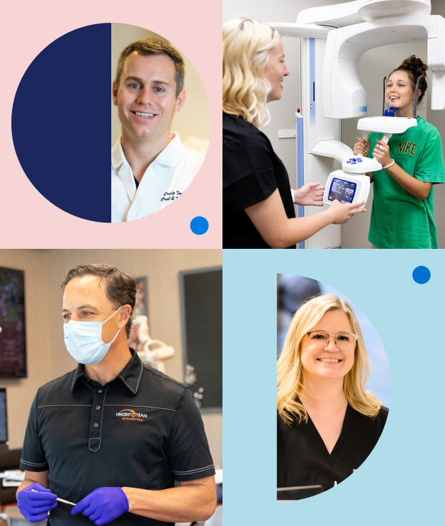
Making Oral Healthcare Easier, More Accessible, and Enjoyable for dental specialists and the communities they care for.
Join the country’s best pediatric dentists, orthodontists, and oral surgeons to grow your practice.
Partner With Us
CASE STUDIES
The Impact of Partnership
Dive into the transformative alliances shaped by SDB through these in-depth case studies.
The Specialty Dental Brands Partnership Model
We provide the most comprehensive and dependable behind-the-scenes support so our specialists can focus on what matters most: their patients.
Maintain clinical autonomy
Support services to help manage your practice
Equity ownership opportunities
New referrals from a multi-specialty network
Continued mentorship opportunities
Integration plan with extended onboarding support
Business Support
Specialty Dental Brands provides best-in-class support services to help you grow your practice and achieve your goals.
Our People Management team supports your practice through talent acquisition, benefits, payroll, and employee performance management.
We offer consistent IT support with a reliable HIPAA-compliant infrastructure, which helps you keep day-to-day operations running smoothly and efficiently.
Our financial services and accounting team helps to maximize your practice revenue by evaluating metrics based on data-driven trends and establishing achievable goals.
Our partners receive personalized monitoring with comprehensive staff compliance programs, ensuring all federal, state, and local mandates are followed.
Our dedicated marketing team helps you reach prospective patients and increase your referral network. We specialize in new patient acquisition, digital and print advertising, referral marketing, and community events.
Committed to excellence, our operational leaders help you develop plans to drive growth and efficiency while handling the oversight of key systems and financial diligence.
Associate Doctors & Residents
Specialty Dental Brands offers a path to partnership and equity for high performing associates and residents.
Start The Conversation
Please fill out the form below


